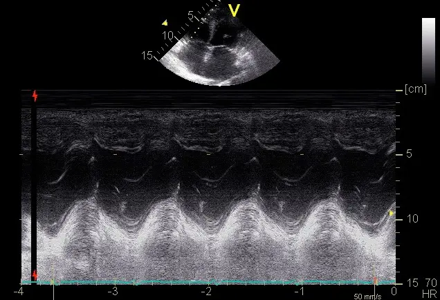
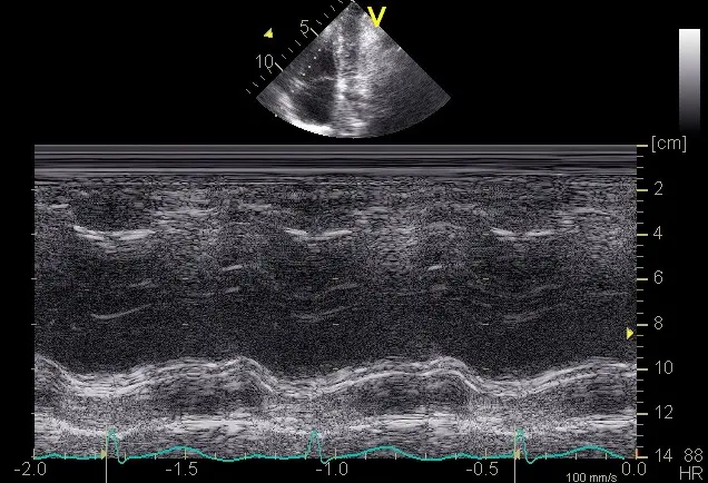
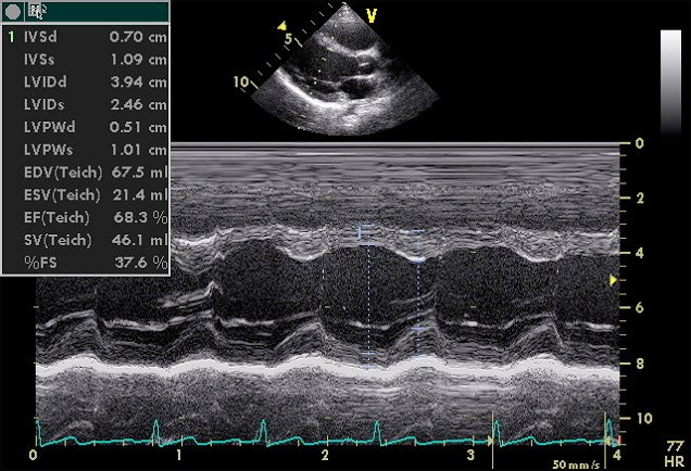
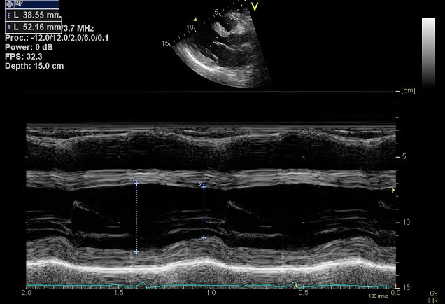
Eleven openly licensed clips, then the SCCM course clips used with permission.
Short teaching loops, not complete examinations.
Heart
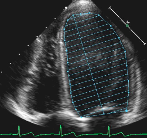
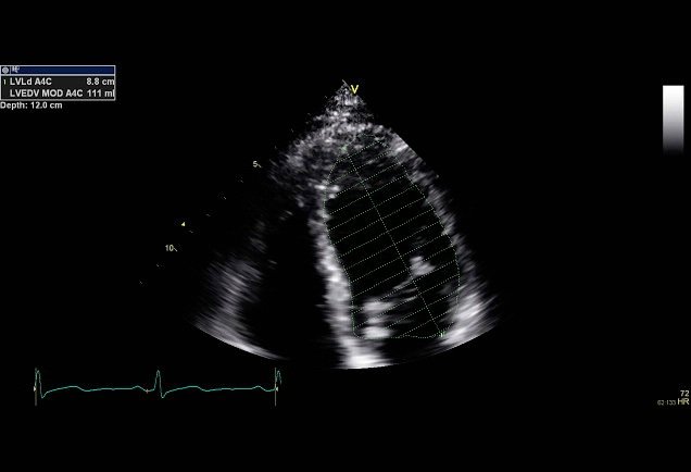
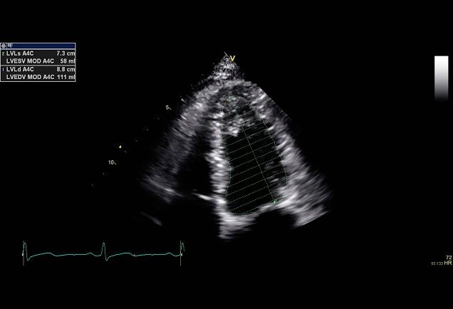
Normal apical four-chamber view
Identify the four chambers, septum and atrioventricular valves across the cardiac cycle.
A single normal example does not exclude cardiac disease or establish an ejection fraction. This short loop is intended for orientation and qualitative motion assessment.
I.A.C. van der Bilt, MD, Department of Echocardiography AMC; CardioNetworks/ECHOpedia (Vdbilt) (2007) · CC BY-SA 3.0
Examination-date area at the upper left masked; original creator credits and scan anatomy retained. Transcoded to H.264 MP4 without changing sequence or playback speed. Audio and source container metadata removed. JPEG poster extracted at 0.15 seconds; it is a still from the processed clinical clip.
This video and its poster remain under CC BY-SA 3.0.
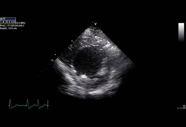
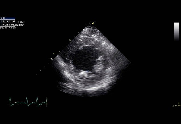
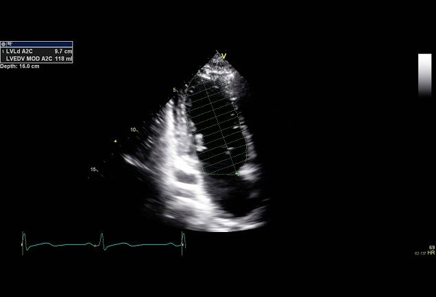
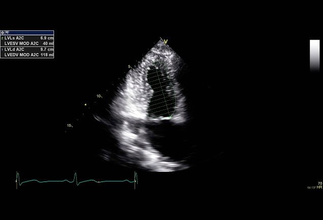
Follow circumferential wall thickening and inward motion at the papillary-muscle level.
Interpret alongside other levels and views. This loop is not a quantitative EF measurement, and foreshortening or obliquity can alter the apparent geometry.
I.A.C. van der Bilt, MD, Department of Echocardiography AMC; CardioNetworks/ECHOpedia (Vdbilt) (2007) · CC BY-SA 3.0
Examination-date area at the upper left masked; original creator credits and scan anatomy retained. Transcoded to H.264 MP4 without changing sequence or playback speed. Audio and source container metadata removed. JPEG poster extracted at 0.15 seconds; it is a still from the processed clinical clip.
This video and its poster remain under CC BY-SA 3.0.
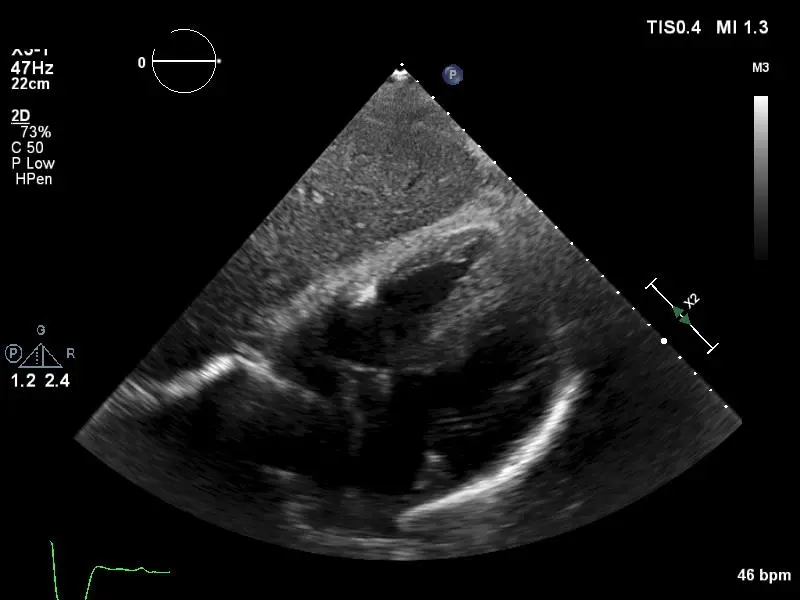
Recognise pericardial fluid and inspect right-sided chamber motion in an apical cine from a published tamponade teaching case.
Confirm collapse timing with ECG or M-mode and integrate haemodynamics. Effusion alone does not diagnose tamponade, and the clip is not an instruction to drain.
Ben Smith, Ultrasound of the Week (2017) · CC BY-SA 4.0
Transcoded to H.264 MP4 without changing sequence or playback speed. Audio and source container metadata removed. JPEG poster extracted at 0.15 seconds; it is a still from the processed clinical clip.
This video and its poster remain under CC BY-SA 4.0.
The identifying/examination header across the top 28 pixels masked outside the scan anatomy. Transcoded to H.264 MP4 without changing sequence or playback speed. Audio and source container metadata removed. JPEG poster extracted at 0.15 seconds; it is a still from the processed clinical clip.
Compare the pleural line with the sliding example. Movement beneath it is markedly reduced or absent.
Absent sliding is nonspecific. Consider apnoea, pleural adhesions, mainstem intubation, severe lung disease and pneumothorax; integrate other signs and clinical context.
Gillman LM, Ball CG, Panebianco N, Al-Kadi A, Kirkpatrick AW (2009) · CC BY 2.0
The identifying/examination header across the top 28 pixels masked outside the scan anatomy. Transcoded to H.264 MP4 without changing sequence or playback speed. Audio and source container metadata removed. JPEG poster extracted at 0.15 seconds; it is a still from the processed clinical clip.
Identify vertical bright artefacts arising at the pleural line and extending through the displayed image.
The original source calls these comet-tail artefacts. Use contemporary B-line definitions and scan settings; the appearance alone does not establish pulmonary oedema or its cause.
Gillman LM, Ball CG, Panebianco N, Al-Kadi A, Kirkpatrick AW (2009) · CC BY 2.0
The identifying/examination header across the top 28 pixels masked outside the scan anatomy. Transcoded to H.264 MP4 without changing sequence or playback speed. Audio and source container metadata removed. JPEG poster extracted at 0.15 seconds; it is a still from the processed clinical clip.
Follow the respiratory transition between sliding lung and a region without sliding in the published pneumothorax example.
Confirm the expected A-line pattern on the non-sliding side and distinguish lung-edge mimics. Absence of a lung point does not exclude pneumothorax; do not infer tension physiology from the clip.
Gillman LM, Ball CG, Panebianco N, Al-Kadi A, Kirkpatrick AW (2009) · CC BY 2.0
The identifying/examination header across the top 28 pixels masked outside the scan anatomy. Transcoded to H.264 MP4 without changing sequence or playback speed. Audio and source container metadata removed. JPEG poster extracted at 0.15 seconds; it is a still from the processed clinical clip.
Sliding lung moves over mediastinal soft tissue with cardiac motion. The non-lung side lacks the expected A-line/no-sliding pattern.
CT excluded pneumothorax in this published case. Compare the entire image pattern with the true lung-point example; a moving boundary alone is insufficient.
Identifying/time header masked; black pillarboxes removed and display proportionally resized from the original 1920 × 1080 container to 720 × 540. Transcoded to H.264 MP4 without changing sequence or playback speed. Audio and source container metadata removed. JPEG poster extracted at 0.15 seconds; it is a still from the processed clinical clip.
The identifying/examination header across the top 28 pixels masked outside the scan anatomy. Transcoded to H.264 MP4 without changing sequence or playback speed. Audio and source container metadata removed. JPEG poster extracted at 0.15 seconds; it is a still from the processed clinical clip.
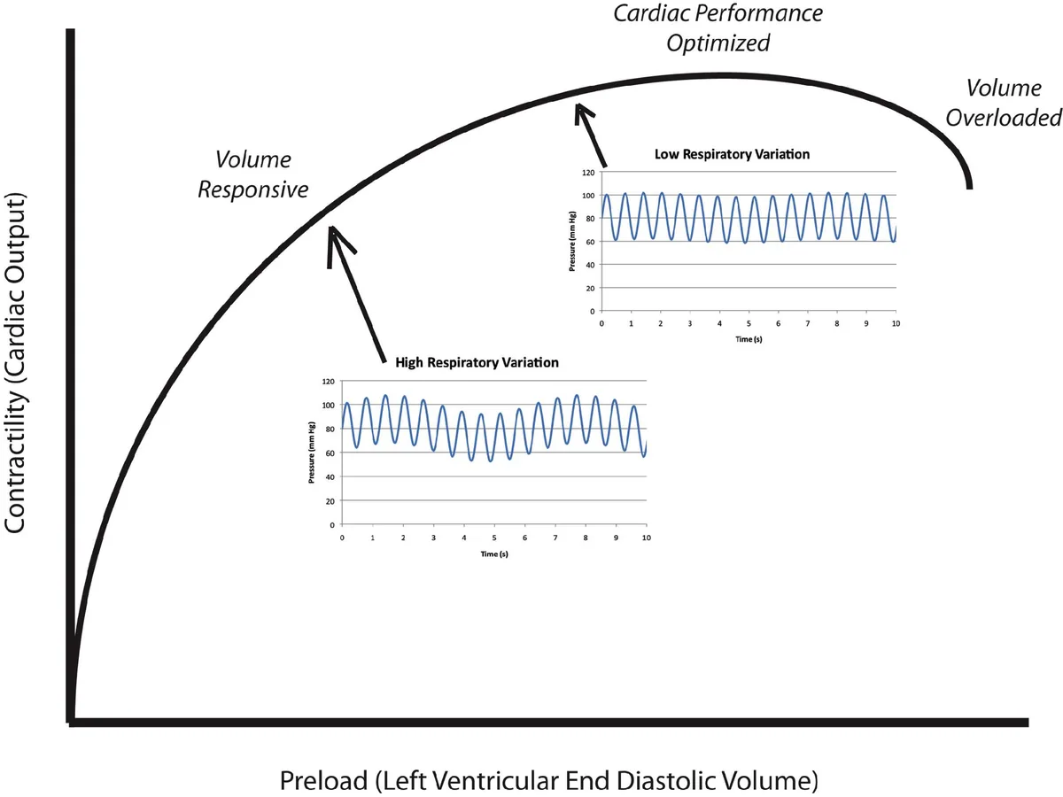
Observe changes in IVC calibre and position during respiration.
IVC variation is influenced by breathing effort, ventilation, abdominal pressure and right-heart function. This clip cannot prove hypovolaemia or predict fluid responsiveness.
Gillman LM, Ball CG, Panebianco N, Al-Kadi A, Kirkpatrick AW (2009) · CC BY 2.0
Non-square pixels normalised to 640 × 480 while preserving the original 4:3 display geometry; the source import already omitted its identifying header. Transcoded to H.264 MP4 without changing sequence or playback speed. Audio and source container metadata removed. JPEG poster extracted at 0.15 seconds; it is a still from the processed clinical clip.
Non-square pixels normalised to 640 × 480 while preserving the original 4:3 display geometry; the source import already omitted its identifying header. Transcoded to H.264 MP4 without changing sequence or playback speed. Audio and source container metadata removed. JPEG poster extracted at 0.15 seconds; it is a still from the processed clinical clip.
From the SCCM Critical Care Ultrasound: Adult course, right-ventricle lecture. Used with permission; not openly licensed, so do not copy them for reuse. Headers cropped and metadata removed; per-file details in media-attributions.json.
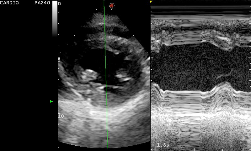
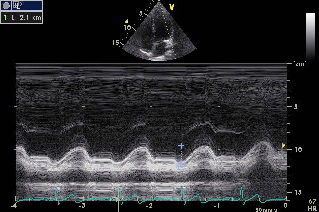
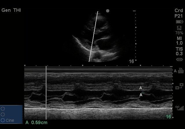
After VF arrest: severe hypokinesis to akinesis of the anterior wall, apex and septum with preserved lateral and inferior walls (course answer: cardiac catheterisation).
No asset-specific open reuse licence verified on the reviewed page. Link to the original.
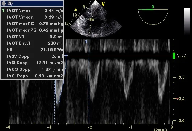
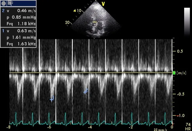
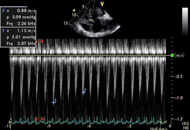
These clinical clips supplement the original interactive diagrams. Source resolution varies. The collection does not yet supply every Doppler or pathological example; use the lecture explanations and references for those topics.
Reuse: each clip and its poster retain the Creative Commons licence stated beside them. These licences take precedence over the website’s general copyright notice. Give the creator, source, licence and changes when reusing a file; preserve the stated ShareAlike licence for adapted CC BY-SA files.